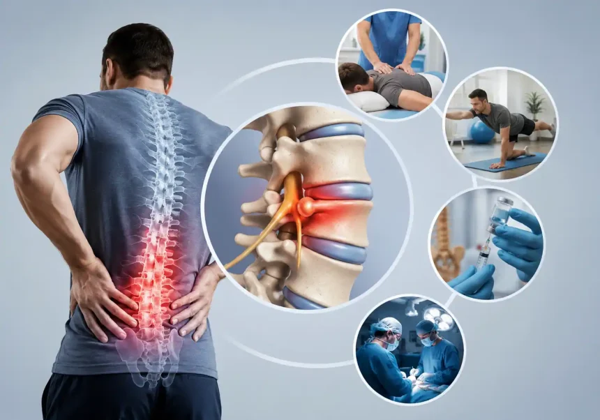
What Is a Herniated Disc?
A herniated disc — also called a slipped disc or ruptured disc — occurs when the soft, gel-like center of a spinal disc pushes through a tear in its tougher outer layer. Think of a spinal disc like a jelly donut. The outer dough (the annulus fibrosus) is firm and fibrous, while the inner jelly (the nucleus pulposus) is soft and cushioning. When that outer layer cracks, the inner material leaks out and can press against nearby nerves, triggering pain, numbness, or weakness.
Herniated discs most commonly occur in the lower back (lumbar spine), though they can also develop in the neck (cervical spine). In rare cases, they appear in the upper back (thoracic spine). The condition affects millions of people worldwide each year, with adults between 30 and 50 being the most commonly impacted group. Men are roughly twice as likely as women to develop a herniated disc.
Recognizing the Symptoms
Lumbar Herniated Disc (Lower Back)
When a disc herniates in the lower back, symptoms often travel beyond the back itself. The hallmark sign is sciatica — a sharp, shooting pain that radiates from the buttock down through the back of the leg and sometimes into the foot. This occurs because the herniated material compresses the sciatic nerve, the largest nerve in the body.
Other common lumbar symptoms include:
- Persistent lower back pain that worsens with sitting, bending, or coughing
- Numbness or tingling in the leg or foot
- Muscle weakness that affects walking or standing on tiptoes
- A burning sensation running along the path of the affected nerve
Cervical Herniated Disc (Neck)
A herniated disc in the neck produces symptoms that radiate into the shoulders, arms, and hands. Patients often report:
- Sharp or dull neck pain that intensifies with certain head positions
- Tingling or numbness radiating down one arm
- Muscle weakness in the biceps, triceps, or hand grip
- Headaches originating at the base of the skull
When to Seek Emergency Care
Certain symptoms signal a medical emergency requiring immediate attention. Cauda equina syndrome, though rare, occurs when the bundle of nerves at the base of the spinal cord is compressed. Red flags include:
- Sudden loss of bladder or bowel control
- Progressive numbness in the saddle area (inner thighs, genitals, buttocks)
- Rapidly worsening leg weakness that impairs the ability to walk
If any of these symptoms appear, head to an emergency room without delay. Early surgical intervention is critical to prevent permanent damage.
How Doctors Diagnose a Herniated Disc
Diagnosis begins with a thorough physical examination. A doctor will assess your range of motion, test muscle strength, check reflexes, and identify areas of tenderness or numbness. The straight leg raise test — where the doctor lifts your leg while you lie flat — is a classic clinical tool for detecting lumbar disc herniation.
If the physical exam suggests a herniated disc, imaging tests provide confirmation:
- Magnetic Resonance Imaging (MRI): The gold standard. An MRI produces detailed images of soft tissues, clearly showing the location and severity of disc herniation and any nerve compression.
- CT Scan: Sometimes used when MRI is contraindicated, such as for patients with certain implants.
- X-rays: Cannot visualize herniated discs directly but help rule out other causes of pain like fractures or arthritis.
- Electromyography (EMG): Measures electrical activity in muscles and can pinpoint which nerve is affected.
Non-Surgical Treatment Options
The vast majority of herniated discs — roughly 80 to 90 percent — resolve within six to twelve weeks without surgical intervention. Most treatment plans begin conservatively and escalate only when necessary.
Rest and Activity Modification
Brief rest can ease acute pain, but staying immobile for more than a day or two often backfires. Gentle movement promotes blood flow and prevents muscle stiffness. Modify activities to avoid heavy lifting, prolonged sitting, and movements that twist the spine.
Physical Therapy
A structured physical therapy program forms the backbone of non-surgical treatment. A physical therapist designs exercises that:
- Strengthen core muscles that support the spine
- Improve flexibility and range of motion
- Teach proper posture and body mechanics during daily activities
- Gently stretch tight muscles like the hamstrings and hip flexors
Consistency matters. Patients who adhere to their home exercise programs tend to recover faster and have lower recurrence rates.
Medications
Pain management during the acute phase may involve:
- Over-the-counter NSAIDs like ibuprofen or naproxen to reduce inflammation
- Muscle relaxants for associated muscle spasms
- Neuropathic pain medications such as gabapentin or pregabalin for nerve-related burning or tingling
- Oral corticosteroids in short courses for severe inflammation
Opioid painkillers are generally avoided due to addiction risks and limited evidence of long-term benefit for disc-related pain.
Epidural Steroid Injections
When oral medications and physical therapy provide insufficient relief, an epidural steroid injection can deliver a potent anti-inflammatory directly to the affected nerve root. Performed under fluoroscopic (X-ray) guidance, the injection can reduce pain for weeks to months, creating a window for physical therapy to work more effectively.
Surgical Treatment Options
Surgery enters the conversation when conservative measures fail after six to twelve weeks, or when neurological deficits worsen. The goal is always to relieve nerve compression while preserving spinal stability.
Microdiscectomy
Microdiscectomy remains the most common surgical procedure for lumbar herniated discs. Using a microscope for magnification, the surgeon makes a small incision and removes only the herniated portion of the disc compressing the nerve. The procedure is minimally invasive, with most patients going home the same day or after a single night in the hospital. Success rates for leg pain relief exceed 90 percent in properly selected patients.
Laminectomy or Laminotomy
These procedures involve removing a portion of the vertebral bone (the lamina) to create more space for the nerves. A laminectomy removes the entire lamina, while a laminotomy removes only part. Both are sometimes performed alongside microdiscectomy.
Artificial Disc Replacement
For certain patients, particularly younger individuals with isolated disc disease, artificial disc replacement offers an alternative to fusion. The damaged disc is removed and replaced with a mechanical device that preserves motion at the affected spinal segment. This approach may reduce the risk of adjacent segment degeneration compared to fusion.
Spinal Fusion
When multiple discs are involved or spinal instability is present, fusion surgery permanently connects two or more vertebrae using bone grafts, screws, and rods. Fusion eliminates motion at the affected segment, which can relieve pain but increases stress on adjacent discs over time.
Recovery and Rehabilitation
The First Two Weeks
Immediately after surgery (or when beginning conservative treatment), focus on gentle walking. Short, frequent walks — starting with five to ten minutes — promote circulation and prevent complications. Avoid bending, lifting anything heavier than a few kilograms, and twisting the spine. Use a grabber tool for picking up objects from the floor.
Weeks Two Through Six
Gradually increase walking duration and begin formal physical therapy. The therapist introduces gentle core stabilization exercises and may use manual therapy techniques to address muscle tension. Return to desk work is often possible during this phase, provided ergonomic seating and frequent standing breaks are used.
Beyond Six Weeks
Most patients return to normal daily activities. Continue core strengthening exercises as a permanent part of your routine — a strong core is your best defense against recurrence. Avoid high-impact activities like running on hard surfaces until cleared by your doctor. Swimming and stationary cycling are excellent low-impact options for cardiovascular fitness.
Prevention Strategies
Recurrence prevention centers on spinal hygiene and lifestyle habits:
- Maintain a healthy weight: Excess body weight, especially around the abdomen, increases stress on the lumbar discs.
- Practice proper lifting technique: Bend at the knees, keep the load close to your body, and avoid twisting while lifting.
- Stay active: Regular low-impact exercise keeps discs nourished, as spinal discs receive nutrients through movement rather than direct blood supply.
- Quit smoking: Smoking accelerates disc degeneration by reducing oxygen and nutrient delivery to spinal tissues.
- Set up an ergonomic workstation: Position your monitor at eye level, keep your feet flat on the floor, and use a chair with proper lumbar support.
Living with a Herniated Disc
A herniated disc diagnosis does not mean a lifetime of pain. Most people recover fully with appropriate treatment and go on to lead active, unrestricted lives. The key is early intervention, patience with the healing process, and commitment to the lifestyle habits that protect your spine for years to come.
If symptoms persist despite conservative care, do not delay seeking a spine specialist’s opinion. Early surgical intervention, when truly needed, produces better outcomes than waiting until nerve damage becomes severe.
Author: Dr. Michael Chen is a board-certified physical medicine and rehabilitation specialist with over twelve years of experience treating spinal conditions.